
If you have ever wondered whether your mammogram is actually catching everything it should, you are not alone. It is one of the most common questions women ask before and after their screening. The honest answer is that mammograms are the best tool we have for finding breast cancer early, but they are not perfect. Understanding what they can and cannot do helps you make smarter decisions about your health.
This guide walks you through everything in plain terms: how the test works, what the accuracy numbers really mean, what to do if you get an abnormal result, and how to figure out the right screening schedule for your age and risk level. We have also included comparison tables and step-by-step guides to help you know exactly what to ask at your next breast health appointment.
Table of Contents
What Is a Mammogram?
A mammogram is a low-dose X-ray of the breast. It is designed to spot abnormalities in breast tissue that are too small or too deep to feel by hand. A radiologist looks at the images for tiny calcium deposits (called microcalcifications) or unusual masses that could be early signs of cancer.
Why Getting Screened Matters
Screening does not prevent breast cancer. What it does is find it early, when it is most treatable. According to the National Cancer Institute’s SEER database, when breast cancer is caught early, before it has spread outside the breast, the 5-year survival rate is 99.3%. That drops to 86.3% when it has reached nearby lymph nodes, and 31% when it has spread to other parts of the body. (NCI: Breast Cancer Survival Rates)
The American Cancer Society reports that women who get regular screenings have a 26% lower breast cancer death rate than women who skip them. (National Breast Cancer Foundation, citing ACS data)
2D vs. 3D Mammograms: What Is the Difference?
There are two main types of mammograms available today. Both use X-rays, but they work differently:
- 2D Mammography: The standard type. It takes two flat X-ray images of each breast, one from the top and one from the side.
- 3D Mammography (also called Digital Breast Tomosynthesis or DBT): This newer technology takes multiple images from different angles and combines them into a 3D view. Radiologists can scroll through the breast tissue layer by layer, like flipping through the pages of a book. Studies show it catches about 29% more cancers than 2D mammography and sends fewer women back for unnecessary follow-up. (Friedewald et al., cited in Clinical Trials DBT Protocol)
Here is a side-by-side look at how the two compare:
| Feature | 2D Mammogram | 3D Mammogram (DBT) |
|---|---|---|
| How it works | Two flat X-ray images per breast | Many images from different angles, combined into 3D layers |
| Cancer detection rate | About 4.2 per 1,000 screenings | About 5.4 per 1,000 screenings (29% higher) |
| Unnecessary callbacks | Higher (overlapping tissue can look suspicious) | Lower (the 3D view reduces confusion) |
| Dense breasts | Misses 52–70% of cancers in extremely dense breasts | Better than 2D, though still not perfect in very dense tissue |
| Radiation | About 0.4 mSv (roughly 7 weeks of everyday background radiation) | Slightly more, about 0.5 to 1.0 mSv. Still very low. |
| Where to get it | Available everywhere | Available at most hospitals and imaging centers in the U.S. |
| Insurance coverage | Covered by most plans | Increasingly covered; small extra fee at some facilities |
| Best for | When 3D is not available | Most women, especially those with dense breasts |
Sources: Friedewald et al., DBT Clinical Trials Protocol; BreastCancer.org; Maurer Foundation, citing ACS
A simple question to ask at your appointment: “Will my mammogram today be 2D or 3D?” If 3D is available at your facility, it is generally the better option.
When Should You Start? 2024 Guidelines Compared
You may have heard different things from different doctors about when to start mammograms. That is because the major medical organizations do not fully agree. Here is where each one stands as of 2024:
| Organization | Start Age | How Often | When to Stop |
|---|---|---|---|
| USPSTF (U.S. Preventive Services Task Force) | 40 | Every 2 years | Age 74 |
| American Cancer Society | Option at 40; strongly recommended by 45 | Annual ages 45 to 54; every 1 to 2 years at 55 and older | As long as you are in good health |
| American College of Radiology (ACR) | 40 | Annual | No upper limit if healthy |
| Women at higher risk | Before 40 in many cases | Annual, often with MRI added | Discuss with your provider |
Sources: USPSTF 2024; American Cancer Society; ACR 2023 Guidelines via BreastCancer.org
The big news in 2024: the USPSTF lowered its recommended start age from 50 to 40. That change alone could reduce breast cancer deaths by about 20%. (USPSTF 2024, JAMA)
If you are not sure which guideline to follow, a good rule of thumb is to start talking to your doctor about mammograms at age 40 and make a plan together based on your personal health history.
What If You Are Under 40?
Routine mammograms are not recommended for most women under 40. The main reason is that younger women tend to have denser breast tissue, which makes mammograms harder to read. Dense tissue and tumors both appear white on an X-ray, so it is harder to tell them apart. This leads to more false alarms and follow-up tests that turn out to be nothing. (Breast Cancer Research Foundation)
That said, some women under 40 should start screening earlier. Talk to your doctor sooner if any of these apply to you:
- You carry a BRCA1 or BRCA2 gene mutation. Most guidelines recommend starting annual mammograms plus annual MRI at age 25 to 30.
- A close relative (mother, sister, or daughter) was diagnosed with breast cancer before age 50. Consider starting screening 10 years before the age your relative was diagnosed.
- You had radiation therapy to the chest (for example, for Hodgkin’s lymphoma). Screening typically starts 8 to 10 years after treatment.
- A prior biopsy found a high-risk breast lesion such as atypical ductal hyperplasia or lobular carcinoma in situ.
- You are of Ashkenazi Jewish descent, which is associated with higher rates of BRCA mutations. The ACR recommends a risk assessment at age 25. (ACR 2024)
If any of these sound like you, ask your OB-GYN about a breast cancer risk assessment. This is a short conversation using a validated scoring tool (such as the Gail model) that can help you and your doctor decide on the right screening plan.
Which Screening Plan Fits You?
Use the table below to find the profile that sounds most like you, then bring it to your next appointment as a starting point for conversation. Your doctor will help you tailor it to your specific situation.
| Your Situation | Suggested Start Age | How Often | Additional Tests to Ask About | Why |
|---|---|---|---|---|
| Average risk, no family history, breasts are not dense | 40 | Every 1 to 2 years | Standard 2D or 3D mammogram | USPSTF 2024 lowered the start age to 40 for all average-risk women |
| Dense breasts (heterogeneous) | 40 | Annual | 3D mammogram plus consider adding an ultrasound | Dense tissue can hide tumors. Adding an ultrasound can find 3 to 4 extra cancers per 1,000 women. |
| Extremely dense breasts | 40 | Annual | 3D mammogram plus ultrasound or abbreviated MRI | Mammogram alone finds only 30 to 48% of cancers in extremely dense breasts. An MRI finds the most. |
| Mother or sister had breast cancer | 10 years before relative’s diagnosis age, or 40, whichever comes first | Annual | Mammogram plus consider MRI depending on your risk score | A close family history roughly doubles your risk |
| BRCA1 or BRCA2 gene mutation | 25 to 30 | Annual mammogram plus annual MRI, alternating every 6 months | MRI is strongly recommended alongside mammogram | Lifetime risk can reach 50 to 85%. MRI catches cancers that mammograms miss in high-risk patients. |
| Black women (any risk level) | 40 (risk assessment at 25 per ACR) | Annual | Ask your provider about a full risk assessment | Black women have higher breast cancer death rates and are more likely to be diagnosed younger and with more aggressive cancers |
| Prior chest radiation therapy | 8 to 10 years after radiation, or age 25, whichever comes later | Annual mammogram plus annual MRI | Both mammogram and MRI are recommended | Chest radiation significantly raises lifetime breast cancer risk |
Sources: USPSTF 2024; BreastCancer.org; Society of Breast Imaging
How to Prepare for Your Mammogram
A few simple steps can make a real difference in how clear your images come out:
- Pick the right time of the month. If you still have periods, try to schedule your mammogram for the week after your period ends. Your breasts tend to be less tender then.
- Skip deodorant and lotion that day. Do not wear deodorant, antiperspirant, powder, or lotion under your arms or on your breasts. The tiny metallic particles in these products can show up on the X-ray as white spots and be mistaken for calcifications.
- Bring your old mammograms. If you have had mammograms at a different facility, bring those images or ask that they be sent ahead. Having past scans for comparison cuts your risk of getting a false alarm by about half. (American Cancer Society)
- Wear a top and separate bottoms. You will only need to undress from the waist up, so a two-piece outfit makes things easier.
- Tell your technologist your full history. Let them know about any breast symptoms, past surgeries, hormone use, or family history of breast cancer. Also mention if you are pregnant or breastfeeding.
Is Mammogram Radiation Dangerous?
This is one of the most common worries women have, and the short answer is no. The amount of radiation in a mammogram is very small.
According to the American Cancer Society, a standard mammogram of both breasts uses about 0.4 mSv of radiation. That is a unit called a millisievert. Here is what that means in everyday terms: (Maurer Foundation, citing ACS)
| Radiation source | Dose |
|---|---|
| Standard mammogram (both breasts) | 0.4 mSv |
| Normal background radiation you get every year just from living (U.S. average) | 3.0 mSv per year |
| How long to get that same 0.4 mSv from everyday life | About 7 weeks |
| A roundtrip flight from New York to Los Angeles | About 0.04 mSv (10 times less than a mammogram) |
| A chest CT scan | About 7 mSv (17 times more than a mammogram) |
| Annual safety limit for radiation workers | 50 mSv (125 times more than a mammogram) |
| 3D mammogram (DBT) | About 0.5 to 1.0 mSv. Slightly more than 2D, still very low. |
The estimated lifetime risk of developing a radiation-induced cancer from a single mammogram is about 5 cases per 100,000 women. The FDA, NCI, and American Cancer Society all agree that the benefit of early detection far outweighs that tiny risk. (Hendrick RE, Tredennick T, PMC 2016)
Mammography radiation is also tightly regulated by federal law. The Mammography Quality Standards Act (MQSA), enforced by the FDA, sets strict dose limits for every facility in the country. (Radiology Ltd.)
Bottom line: Skipping a mammogram because of radiation fear is a far bigger health risk than the scan itself.
How Accurate Are Mammograms?
No medical test is 100% accurate, and mammograms are no exception. But understanding what “accurate” really means will help you put your results in context.
What the Numbers Actually Mean
Doctors measure mammogram accuracy in two ways:
- Sensitivity is the test’s ability to find cancer when it is there. A higher number is better.
- Specificity is the test’s ability to come back normal when there is no cancer. A higher number means fewer false alarms.
On average, mammograms find cancer in about 87 out of every 100 women who actually have it. That means about 13 out of 100 cancers are missed. The test correctly identifies healthy tissue as healthy about 90 to 95% of the time, meaning 5 to 10 women out of 100 without cancer will still get called back for more tests.
Those numbers can change a lot depending on your breast type. In women with fatty breasts, mammography catches up to 98% of cancers. In women with extremely dense breasts, it drops to just 30 to 48%. (BreastCancer.org)
False Positives: When You Get Called Back But Do Not Have Cancer
A false positive is when your mammogram looks like something might be wrong, but after more testing, it turns out to be nothing. This is very common. Here is what the data shows:
- About 1 in 10 mammograms leads to a callback for more imaging. Of those callbacks, only about 7% actually turn out to be cancer. The other 93% are false alarms. (National Cancer Institute, 2024)
- Women in their 40s get more false positives: 10 to 12% of mammograms in that age group lead to a callback. (ASCO Post, 2024)
- If you get annual mammograms for 10 years, there is a 50 to 60% chance you will get at least one false alarm at some point. About 7 to 12% of women will have a false alarm serious enough to lead to a biopsy recommendation. (ASCO Post, 2024)
- The American Cancer Society says false positives are more common in younger women, women with dense breasts, women who have had a breast biopsy before, women with a family history of breast cancer, and women who take estrogen. (ACS)
One important thing to know: a 2024 study from the National Cancer Institute looked at 3.5 million mammograms and found that women who got a false alarm were less likely to come back for future screenings. Only 61% returned, compared to 77% of women who had a normal result. (NCI, 2024) If you get a callback, please do not let it scare you away from future screenings. Talk to your doctor about what it means.
False Negatives: When Cancer Gets Missed
A false negative is the opposite: your mammogram looks normal, but cancer is actually present. According to the National Cancer Institute, mammograms miss about 1 in 8 breast cancers. This happens more often in women with dense breasts because the dense tissue can hide a tumor on the X-ray. (NCI)
This is why a normal mammogram result does not mean you should stop paying attention to your breasts. If you notice a lump, pain, skin changes, or any other new symptom, contact your doctor right away even if your last mammogram was clear.
What Affects Mammogram Accuracy
- Breast density. The single biggest factor. Dense breasts contain more fibrous and glandular tissue. On an X-ray, dense tissue and tumors both appear white, so they can be hard to tell apart. About 40% of women over 40 have dense breasts. Since September 2024, all U.S. mammography facilities are required by federal law to tell you if your breasts are dense. (NCI; FDA MQSA 2024)
- Age. Younger women tend to have denser breasts, which lowers accuracy. As you get older, breast tissue typically becomes fattier and easier to read.
- How often you screen. Annual mammograms catch more cancers than screenings every two years. But more frequent screening also increases the chance of a false alarm over time.
- Having past mammograms to compare. When a radiologist can compare your current images to older ones, your risk of a false alarm drops by about half. This is one of the best reasons to stay with the same facility year after year. (ACS)
- The radiologist reading your scan. Experience and how many mammograms a radiologist reads each year both affect accuracy.
- The type of equipment. 3D mammography is more accurate than 2D and leads to fewer unnecessary callbacks, especially for women with dense breasts.
- AI assistance. Artificial intelligence tools are now used at many facilities to help radiologists spot suspicious areas. See the section below for what the research says.
How AI Is Changing Mammogram Accuracy
Artificial intelligence is becoming a real tool in mammography, not just a buzzword. As of mid-2025, the FDA had cleared nearly 900 radiology AI tools, with a growing number designed specifically for mammograms. (Intuition Labs, 2025)
The most compelling evidence comes from a large study in Sweden called the MASAI trial. It found that AI-assisted mammogram reading:
- Found 29% more cancers than standard radiologist reading alone
- Reduced cancers missed between screenings by 19%
- Detected 6.4 cancers per 1,000 screenings versus 5.0 without AI (CancerNetwork, 2026)
AI tools are trained on hundreds of thousands of mammogram images. They flag areas that look suspicious, help prioritize urgent cases, and can act as a second set of eyes in facilities where two radiologists are not available.
There are some cautions worth knowing. Most of the big AI studies were done in Europe, and researchers are still studying whether the results hold up equally well across the more diverse U.S. population. The Susan G. Komen Foundation notes that “it is unknown whether these results will be the same in the U.S. or will benefit everyone in the diverse U.S. population.” (Susan G. Komen)
The USPSTF also flagged AI as something to watch carefully, noting that an earlier generation of computer-aided detection tools was adopted widely before studies revealed it sometimes made things worse. (AuntMinnie, 2024)
What you can do: It is fine to ask your screening facility whether they use FDA-cleared AI tools in their mammogram reading process. This is a reasonable question and the answer may affect how confident you feel in your results.
Breast Density: Do Dense Breasts Need Additional Testing?
If you have been told you have dense breasts, a mammogram alone may not give you the full picture. There are several additional tests your doctor might recommend. Here is how they compare:
| Test | Extra Cancers Found per 1,000 Women | False Alarm Risk | Cost and Access | Best For |
|---|---|---|---|---|
| 3D Mammogram (DBT) only | About 1.2 more than 2D | Fewer callbacks than 2D | Widely available; usually covered | All women with dense breasts as a first upgrade |
| Mammogram plus handheld ultrasound | About 3 to 4 extra | Higher; biopsy rate about 2 to 5% | Moderate cost; widely available | Women with moderately dense breasts at average or intermediate risk |
| Mammogram plus automated whole-breast ultrasound (ABUS) | About 2.5 extra | Higher false alarm biopsy rate | Moderate cost; availability is growing | Average-risk women with dense breasts; no technologist needed to hold the probe |
| Mammogram plus abbreviated MRI | About 25.7 extra per 1,000 (the highest of any option) | Detects the smallest tumors (average 9.5 mm) but lower biopsy success rate | Most expensive; not always covered for average-risk women | Extremely dense breasts or intermediate-to-high risk |
| Mammogram plus contrast-enhanced mammography (CEM) | Similar to abbreviated MRI | Similar to abbreviated MRI | Available at some centers; emerging option | Women who cannot have an MRI, such as those with metal implants or claustrophobia |
Sources: BreastCancer.org; Hussein and Abbas et al., PMC 2023; Society of Breast Imaging
A good question to ask your provider: “Are my breasts heterogeneously dense or extremely dense? And which extra test makes sense for my risk level and insurance plan?” The answer is different depending on which category you are in.
The Emotional and Financial Side of False Positive and False Negative Results
A false alarm is stressful. The anxiety of waiting for follow-up results, the extra appointments, and sometimes a biopsy are all real burdens. There are financial costs too. It is completely normal to feel shaken by the experience.
What matters is not letting a false alarm keep you away from future screenings. Research shows that women who get a false positive are less likely to come back for their next mammogram, and that gap in screening is where cancers can grow undetected. (NCI, 2024)
On the other side, a false negative, a clean result when cancer is actually present, can give you a false sense of security. This is why it is important to keep doing regular self-checks and to report any new changes in your breast to your doctor even if your last mammogram was normal.
Why Mammograms Save Lives
Even with their limitations, mammograms have made a dramatic difference in breast cancer outcomes. The evidence is hard to argue with.
The Numbers on Breast Cancer Deaths
Breast cancer deaths in the United States have dropped by 44% since 1989. A large part of that improvement is credited to better screening and earlier detection, alongside advances in treatment. (National Breast Cancer Foundation, citing ACS 2024)
The American Cancer Society estimates that mammography has contributed to about a 40% reduction in breast cancer deaths since 1990. Annual screening, compared to every-two-years screening, has been shown to produce an even greater reduction. (Radiology Ltd., summarizing ACS and ACR data)
The USPSTF’s 2024 research found that starting mammograms at age 40 instead of 50 could prevent 1.3 additional breast cancer deaths per 1,000 women screened, and up to 1.8 per 1,000 among Black women, who face higher mortality rates even at the same stage of diagnosis. (AuntMinnie, 2024)
Finding Cancer Before You Can Feel It
One of the biggest advantages of mammography is catching cancer before you or your doctor can feel a lump. A 3D mammogram can detect cancer up to three years before it becomes palpable (able to be felt). It can also find early-stage cancers like ductal carcinoma in situ (DCIS), a type of cancer that has not yet spread beyond the milk ducts. (Radiology Ltd.)
According to the National Cancer Institute’s SEER data, about 64% of breast cancers are currently diagnosed at the earliest stage, when the 5-year survival rate is 99.3% to 100%. Without regular screening, many of those would be found much later. (NCI SEER)
How Early Detection Changes Your Treatment Options
Finding cancer early does more than improve survival. It also changes what treatment looks like. When cancer is small and caught at an early stage, women are more likely to be candidates for a lumpectomy (removing just the tumor) rather than a full mastectomy (removing the whole breast). They may also be less likely to need aggressive chemotherapy or extensive radiation.
Catching it early generally means a shorter treatment process, fewer side effects, better cosmetic results, and a faster return to normal life.
Common Concerns About Mammograms, Answered
One concern you may have read about is overdiagnosis. This means that sometimes mammograms find slow-growing cancers that might never have caused a problem during a person’s lifetime, and those cancers then get treated unnecessarily.
This is a real issue that researchers are actively studying. But the current medical consensus is that the benefit of catching dangerous, fast-growing cancers early outweighs the risk of over-treating slow ones. The goal going forward is to use better technology and smarter screening to tell the difference between the two more reliably.
Understanding Your Mammogram Results
After your mammogram, a radiologist reviews the images and sends a report to your doctor. Every report uses the same scoring system, called BI-RADS, so that results mean the same thing at every facility in the country.
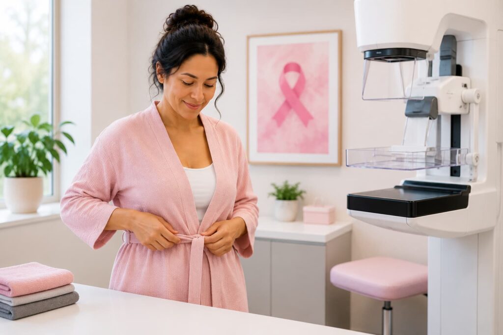
Who Reads Your Mammogram?
A radiologist is a medical doctor who specializes in reading imaging tests. They are trained to spot subtle changes in breast tissue that might signal something worth investigating. Their report tells your primary care doctor or OB-GYN what they found and what to do next.
What Your BI-RADS Score Means
Your result will include a number from 0 to 6. Here is what each one means in plain terms:
- Category 0: The images were not clear enough to make a final call. You will need additional images or an ultrasound before getting a result.
- Category 1: Normal. Nothing to report. Keep up with routine screening.
- Category 2: Normal with a benign finding. The radiologist noticed something, like a cyst, but it is not cancer. Keep up with routine screening.
- Category 3: Probably fine. There is a 98% or higher chance this finding is not cancer. A follow-up scan in about 6 months is recommended to make sure nothing changes.
- Category 4: Needs a closer look. Something is suspicious enough to recommend a biopsy. This category is split into 4A (low suspicion), 4B (moderate), and 4C (high) to give your doctor more detail.
- Category 5: Very likely cancer. There is at least a 95% chance this is cancer. A biopsy is strongly recommended as soon as possible.
- Category 6: Confirmed cancer. This category is only used after a biopsy has already confirmed cancer. Imaging at this stage is typically used to monitor how treatment is working.
Your False Positive Risk Over Time
One of the most common questions women ask is: “What are the chances I will ever get a false alarm?” Here is a breakdown based on published research:
| Years of Annual Screening | Chance of At Least One False Alarm (callback for imaging) | Chance of a False Alarm Leading to Biopsy | What This Means in Practice |
|---|---|---|---|
| 1 year | About 10% | About 1 to 2% | 1 in 10 women gets called back after any single mammogram |
| 5 years | About 35 to 40% | About 5 to 7% | By year 5, nearly 1 in 3 women will have had at least one callback |
| 10 years | About 50 to 60% | About 7 to 12% | More likely than not that you will have had at least one false alarm |
| 10 years (every 2 years instead of annually) | Lower overall | Lower overall | Screening less often reduces false alarms, but means some cancers may be caught later |
| Your risk is higher if you have: | Dense breasts, age under 50, a prior breast biopsy, a family history of breast cancer, estrogen therapy, or if this is your first mammogram with no old ones for comparison | ||
Sources: ASCO Post, 2024; American Cancer Society; Elmore et al., NEJM
The most important thing to remember: Getting called back does not mean you have cancer. It means the radiologist wants a closer look. Most callbacks turn out to be nothing.
What Happens After an Abnormal Result
If your BI-RADS score is 0, 3, 4, or 5, your doctor will walk you through the next steps. Depending on your result, that might be a repeat mammogram with different angles, an ultrasound, an MRI, or a biopsy. Try to remember that most follow-up tests do not end in a cancer diagnosis. Being anxious is completely normal. Staying in close contact with your care team makes the process easier.
Questions to Ask Your Doctor
You are your own best advocate. Here are some questions worth asking at your next appointment:
- What is my BI-RADS score, and what does it mean for me personally?
- Do I have dense breasts? If so, how dense are they, and do I need an additional test?
- Given my age, family history, and other risk factors, which screening schedule makes the most sense for me?
- Should I have a formal breast cancer risk assessment to see if I qualify for MRI screening?
- What are the next steps, and what are the pros and cons of each option?
- If I do get a false alarm, how will we decide together whether to keep doing annual screenings?
Regular mammograms, combined with knowing your own body and staying in conversation with your doctor, give you the strongest possible defense against breast cancer. The evidence from the American Cancer Society, the National Cancer Institute, and decades of research all points to the same conclusion: screening saves lives. Start at 40, stay consistent, and do not be afraid to ask questions.